


Veterinary Diagnostic Imaging For Dogs
As AAHA Accredited Practices, all of our radiology images are submitted to a board certified Veterinary Radiologist for review to assure nothing is missed. This extra step ensures your dog receives the benefit of a specialist every time they have radiographs performed in our practice. Diagnostic imaging includes radiographs (X-rays) and ultrasound, both of which are used as diagnostic tools to collect information on your dog's health. The vast majority of imaging is non-invasive and completely painless. However, some imaging may require sedation because the dog must be still to allow for adequate images to be produced. We use these images to collect information on your dog to help make a medical and sometimes surgical plan.
When Veterinary Diagnostic Imaging Is Necessary For Your Dog
After examining your dog, we may need to collect more information that will lead to a diagnosis and treatment plan. Radiographs are usually the first line of imaging. This may lead to the diagnosis. However, sometimes the next step may be ultrasound to obtain a more thorough view of a particular area of the body.
For instance, if your dog is vomiting and feeling ill, a radiograph will look for possible causes such as obstruction of intestines or an obvious foreign body. They may show some signs of intestinal obstruction, however, before proceeding to surgery, it would be prudent, in some cases, to follow with an abdominal ultrasound. The ultrasound will give more detail of the area and allow more confidence in the treatment plan to move forward with surgery. Occasionally, radiographs and ultrasound allow for a definitive diagnosis but other times they will simply add more information to help put the puzzle together for the best treatment plan for your dog.
As AAHA Accredited practices, Northern Oaks Bird & Animal Hospital and Heritage Oaks Animal Hospital send all of our radiographs to a board certified Veterinary Radiologist for further review.
The four types of Veterinary Diagnostic Imaging our veterinarians may utilize to assist in the diagnosis of your dog's condition are:
- Radiographs
- Ultrasounds (Both In Hospital and Referral)
- MRIs (Referral)
- CT Scans (Referral)
More information on each of these types of imaging is provided below.
Dog Radiographs
Dog Radiographs have been in use throughout the medical community for many decades. Radiographs are by far the most regularly used form of diagnostic imaging in the veterinary industry because they are cost-effective (comparatively speaking), and they can accurately diagnose the state of skeletal structure and composition, large body cavities, and the presence of many foreign objects. Radiographs are totally painless, but some dogs benefit from sedation to reduce anxiety and stress.
Dog radiographs have traditionally been captured on actual film, and still can be when necessary. However, our images are digital which allows us to capture the images on a secure server that our veterinarians can access at any time, and also share with specialists and veterinary radiologists.
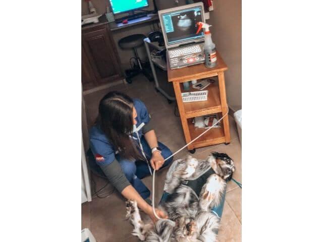
Dog Ultrasounds
A dog ultrasound is the second most common type of diagnostic imaging tool we use to diagnose a dog's medical condition. Ultrasounds use soundwaves to examine and photograph internal tissues in real-time. An ultrasound allows a us to see into a dog's body, allowing for viewing of organs from different angles that are not easily achieved through radiographs. Various organ functions can be observed to determine if they are malfunctioning.
Common symptoms that may cause us to use ultrasound include vomiting, weight loss, kidney impairment or blockage and heart disease.
Dog MRI
Magnetic resonance imaging, or MRI, is the newest form of diagnostic imaging being used for both human and veterinary medicine. Dog MRI equipment generates a very powerful magnetic field, resulting in detailed anatomic images of whatever part of a dog's body is being scanned. No x-rays are involved, and a dog MRI is considered extremely safe. We may recommend this procedure for certain issues. This procedure requires referral to a specialty center.
CT Scans For Dogs
CT scans for dogs, also known as "cat scans," are computer enhanced dog x-ray procedures most often used to evaluate complex parts of the body, such as the head, chest, some joints, and various internal organs. CT scans produce more detailed images than x-rays. In rare instances an MRI may be recommended. This procedure requires referral to a specialty facility.
How Dog Radiographs Influence Veterinary Recommendations
The goal of dog radiographs is to ascertain a diagnosis or obtain a final answer without having to perform further more invasive tests or procedures. For example, a radiograph might show evidence of a tumor of the spine and possibly involve the surrounding muscle. The addition of an MRI would reveal the specific tumor and the extent that the tumor extends into the surrounding muscle tissue. This type of information is very important for a prognosis and treatment plan.
Veterinary diagnostic imaging offers an array of incredibly useful tools within a veterinarian's toolkit. Sometimes a diagnostic imaging session can lead to the need for further diagnostics.